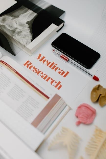
Welcome to the laboratory manual for human anatomy and physiology. This guide provides a comprehensive overview of lab exercises, safety protocols, and essential skills to enhance your learning experience.
1.1 Importance of Anatomy and Physiology in Laboratory Studies
Anatomy and physiology form the foundation of laboratory studies, enabling students to explore the structure and function of the human body. These disciplines provide essential insights into how systems interact, making them vital for understanding complex biological processes. Laboratory work allows for hands-on experience, reinforcing theoretical knowledge through practical observation and experimentation. By examining tissues, cells, and organs, students develop critical thinking and problem-solving skills, which are indispensable in scientific research and clinical practice. The study of anatomy and physiology also fosters a deeper appreciation for human health and disease mechanisms, preparing learners for careers in medicine, research, and healthcare. Through lab investigations, students cultivate precision, accuracy, and analytical abilities, all of which are crucial for advancing medical knowledge and improving patient care. These skills are integral to conducting rigorous scientific inquiries and applying evidence-based practices in real-world scenarios.
1.2 Structure and Organization of the Manual
This laboratory manual is meticulously organized to provide a logical and comprehensive learning experience. It begins with an introduction to the importance of anatomy and physiology, followed by essential skills for lab success. The manual is divided into sections, each focusing on specific body systems, such as skeletal, muscular, circulatory, and nervous systems, among others. Each section includes detailed exercises, step-by-step instructions, and visual aids to enhance understanding. The content is structured to build knowledge progressively, starting with cellular structure and advancing to complex system interactions. Additionally, the manual incorporates safety protocols, waste disposal guidelines, and emergency procedures to ensure a safe learning environment. Tools and resources, such as microscopy techniques and digital simulations, are highlighted to support effective learning. The clear organization of this manual ensures that students can navigate easily, fostering a deeper understanding of human anatomy and physiology through practical exploration and critical thinking.
1.3 Essential Skills for Success in the Lab
Success in the anatomy and physiology lab requires a combination of technical, observational, and critical thinking skills. Students must develop strong dissection techniques to carefully examine specimens and identify structures accurately. Proficiency in microscopy is essential for analyzing cellular and tissue samples, ensuring proper slide preparation and focusing techniques. Measurement and recording skills are critical for collecting accurate data during experiments. Attention to detail is vital to differentiate between similar structures and interpret results effectively. Additionally, effective communication and teamwork skills are necessary for collaborating on group exercises and presenting findings. Time management is also important to complete tasks efficiently within lab sessions. Finally, a curious and analytical mindset fosters deeper understanding and problem-solving in complex anatomical and physiological investigations. Mastering these skills will enhance learning outcomes and prepare students for advanced studies in healthcare and scientific fields.
Safety Protocols and Laboratory Setup
Adhering to safety protocols is crucial in anatomy labs to prevent accidents and ensure a safe learning environment. Proper setup, handling of materials, waste disposal, and emergency preparedness are essential for a well-managed lab.
2.1 General Safety Protocols in the Anatomy Lab
Adherence to safety protocols is paramount in the anatomy lab to minimize risks and ensure a secure environment for all participants. Students must wear appropriate personal protective equipment (PPE), such as gloves, lab coats, and eye protection, when handling specimens or chemicals. Proper handwashing before and after handling materials is essential to prevent contamination. Sharp instruments, like scalpels or forceps, should be used with caution, and students should avoid distractions while working with potentially hazardous tools. The lab should be maintained in a clean and organized state, with clear walkways to prevent accidents. Chemicals and biological materials must be handled according to established guidelines to avoid exposure. Familiarity with emergency equipment, such as eyewash stations and fire extinguishers, is crucial. Strict adherence to these protocols ensures a safe and effective learning experience for everyone involved.
2.2 Handling Biological Specimens and Materials
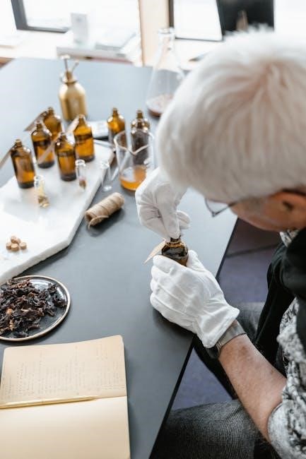
Proper handling of biological specimens and materials is critical to maintaining safety, integrity, and ethical standards in the anatomy lab. Students should always wear gloves when touching specimens to prevent direct contact with potentially hazardous biological materials. Specimens should be handled gently to avoid damage, and tools such as forceps or tongs should be used when appropriate. Biological materials must be stored in designated areas at the correct temperatures to preserve their condition. Labeling and documentation are essential to ensure specimens are easily identifiable and properly organized. After use, specimens should be returned to their storage containers or disposed of according to lab guidelines. Disposal methods must comply with environmental and health regulations to minimize contamination risks. Proper handling techniques not only ensure the quality of the specimens but also contribute to a safe and efficient learning environment.
2.3 Waste Disposal and Environmental Considerations
Proper waste disposal and environmental considerations are essential in the anatomy lab to minimize ecological impact and ensure compliance with regulations. Biological waste, including anatomical specimens and contaminated materials, must be disposed of in designated biohazard containers. Chemical waste should be segregated based on its type and disposed of according to institutional guidelines. Recyclable materials, such as paper and plastic, should be separated from general waste to promote sustainability. All waste containers must be properly labeled to avoid confusion and ensure safe handling. Students should adhere to local and institutional environmental policies to reduce contamination risks. Regular training on waste management practices is crucial to maintain a responsible and eco-friendly laboratory environment. Proper disposal practices not only protect the environment but also contribute to a safe and hygienic workspace for everyone involved.
2.4 Emergency Procedures and First Aid
Emergency procedures and first aid are critical components of laboratory safety. In case of a fire, the RACE acronym should be followed: Rescue anyone in immediate danger, Activate the fire alarm, Contain the fire if possible, and Evacuate the area. Fire extinguishers should be used with the PASS method: Pull the safety pin, Aim the nozzle, Squeeze the handle, and Sweep the extinguishing agent. For chemical spills, evacuate the area and contact trained personnel. First aid kits are available for minor injuries such as cuts or burns, and staff should be trained in their use. In case of eye exposure to chemicals, immediately flush with water for at least 15 minutes; For severe injuries or medical emergencies, call for professional assistance immediately. Regular drills and training ensure preparedness for emergencies, safeguarding both students and staff in the laboratory environment. Proper implementation of these procedures minimizes risks and ensures a prompt response to unexpected situations.
Laboratory Exercises and Investigations
This section provides detailed exercises and investigations to explore human anatomy and physiology through hands-on activities, microscope use, and practical experiments, ensuring a comprehensive understanding of bodily systems and functions.
3.1 Exercise 1: Exploring Cellular Structure
In this exercise, students will examine the basic structure of human cells using a microscope. The activity begins with an introduction to the cell as the fundamental unit of life, emphasizing its importance in anatomy and physiology. Participants will prepare and observe slides of human cells, identifying key components such as the cell membrane, cytoplasm, nucleus, and organelles. The exercise focuses on distinguishing between different cell types, including epithelial, connective, and muscle cells, to understand their specialized functions. Through hands-on observation and guided discussion, students will learn how cellular structures relate to overall bodily functions. This foundational exercise prepares learners for more complex investigations into tissues and systems. Materials required include prepared microscope slides, staining solutions, and a compound microscope. By the end of the exercise, students will be able to identify and describe cellular structures and their roles in maintaining human health.
3.2 Exercise 2: Identifying Types of Tissues
This exercise introduces students to the four primary types of human tissues: epithelial, connective, muscle, and nervous. Participants will learn to distinguish these tissues based on their structural and functional characteristics. Epithelial tissue, which forms linings and coverings, will be examined in samples such as skin or intestinal lining. Connective tissue, including bone, cartilage, and blood, will be studied for its supportive and connective roles. Muscle tissue, both skeletal and smooth, will be observed to understand its role in movement. Nervous tissue, comprising neurons and glial cells, will be explored to highlight its function in communication and control. Using microscopy, students will analyze prepared slides to identify and describe tissue types, reinforcing their understanding of tissue organization and specialization. This exercise builds on cellular structure knowledge, linking it to the hierarchical organization of tissues in the human body.
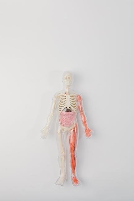
3.3 Exercise 3: Skeletal System Exploration
This exercise provides hands-on experience with the skeletal system, focusing on its structure, classification, and functions. Students will examine human bones and skeletal models to identify major bones, such as the femur, humerus, and skull. The activity emphasizes understanding the axial and appendicular skeleton, including the vertebral column, ribcage, and limb bones. Participants will learn to recognize anatomical features like joints, foramina, and processes, which are critical for bone identification. The exercise also involves assembling a skeletal model to visualize how bones articulate and form the framework of the body. By exploring the skeletal system, students gain insights into its roles in support, protection, and movement. This practical exercise reinforces anatomical terminology and prepares learners for advanced studies in human anatomy and physiology.
3.4 Exercise 4: Muscular System Analysis

This exercise focuses on the study of the muscular system, emphasizing its structure, function, and classification. Students will explore the three types of muscles—skeletal, smooth, and cardiac—through detailed diagrams, models, and human specimens. The activity includes identifying major muscle groups, such as the pectoralis major, quadriceps, and biceps brachii, and understanding their attachments and actions. Participants will also examine the role of tendons and ligaments in muscle function. Practical tasks involve palpating muscles on the human body and assembling a muscular system model to visualize how muscles interact with bones and other tissues. This exercise reinforces the importance of the muscular system in movement, support, and maintaining posture. By analyzing muscle anatomy, students develop a deeper understanding of how the muscular system contributes to overall bodily functions and integrates with other systems like the skeletal and nervous systems.
3.5 Exercise 5: Circulatory System Examination
This exercise focuses on the anatomy and physiology of the circulatory system, often referred to as the cardiovascular system. Students will examine the structural and functional components, including the heart, arteries, veins, and capillaries. The activity involves dissecting animal hearts to identify chambers, valves, and blood vessels, as well as observing blood smears under a microscope to analyze blood cells. Participants will also explore the differences between arterial and venous blood and their roles in oxygenation and nutrient delivery. Practical tasks include tracing blood flow through the heart and understanding the concept of blood pressure regulation. This exercise provides a comprehensive understanding of how the circulatory system maintains homeostasis and supports other bodily systems. By analyzing specimens and models, students gain hands-on experience in identifying key structures and understanding their functions.
3.6 Exercise 6: Respiratory System Investigation
This exercise delves into the anatomy and physiology of the respiratory system, focusing on its role in gas exchange and oxygen delivery to the body. Students will examine the structural components, including the nasal cavity, trachea, bronchi, and lungs, through dissection and microscopy. Activities include identifying the histological features of respiratory tissues, such as alveoli and bronchioles, and understanding the mechanism of breathing. Practical components involve measuring respiratory rates and simulating lung function using models. This exercise also explores the relationship between the respiratory system and other systems, such as the circulatory system, in maintaining homeostasis. By analyzing specimens and conducting experiments, students gain a deeper understanding of how the respiratory system adapts to environmental changes and maintains proper oxygenation of the body. This investigation emphasizes the importance of respiratory health and its impact on overall physiological function.
3.7 Exercise 7: Digestive System Exploration
This exercise focuses on the anatomy and physiology of the digestive system, emphasizing its role in nutrient absorption and waste elimination. Students will explore the structural and functional components, from the oral cavity to the large intestine, using anatomical models and histological slides. Key activities include identifying the layers of the gastrointestinal wall, observing the histological features of different digestive organs, and understanding the processes of mechanical and chemical digestion. Practical tasks involve simulating enzymatic digestion and analyzing the role of accessory organs like the liver and pancreas. The exercise also covers the importance of peristalsis and the absorption of nutrients in the small intestine. By examining the digestive system in detail, students gain insights into how it processes food, maintains gut health, and contributes to overall metabolic function. This investigation highlights the intricate mechanisms that enable the body to extract energy and sustain life.
3.8 Exercise 8: Nervous System Examination
This exercise delves into the structure and function of the nervous system, focusing on its role in controlling body functions and enabling communication between different body parts. Students will examine the central nervous system (CNS), including the brain and spinal cord, and the peripheral nervous system (PNS), comprising nerves and ganglia. Key activities include identifying neural structures using anatomical models and histological slides, analyzing reflex arcs, and exploring the histological features of neurons and glial cells. Practical tasks involve simulating nerve impulse transmission and studying the organization of sensory and motor pathways. The exercise also covers the clinical significance of nervous system structures, such as the impact of nerve damage on motor and sensory functions. By investigating the nervous system, students gain a deeper understanding of its critical role in maintaining homeostasis, regulating responses, and enabling complex cognitive processes. This exploration enhances their appreciation of the nervous system’s intricate mechanisms.
3.9 Exercise 9: Urinary System Analysis
This exercise focuses on the urinary system, which plays a crucial role in eliminating waste, regulating electrolytes, and maintaining fluid balance. Students will examine the structural and functional components of the system, including the kidneys, ureters, bladder, and urethra. Activities involve identifying key anatomical features using models and histological slides, observing the process of urine formation, and understanding the role of nephrons in filtration. Practical tasks include dissecting a pig kidney to visualize renal structures and using microscopy to examine kidney tissue. The exercise also explores the clinical significance of urinary system disorders, such as kidney stones or infections, and their impact on overall health. By analyzing the urinary system, students gain insights into its vital functions, including waste removal, blood pressure regulation, and pH balance. This hands-on investigation enhances understanding of the system’s role in maintaining homeostasis and its interconnection with other body systems.
3.10 Exercise 10: Reproductive System Investigation

This exercise delves into the reproductive system, focusing on its role in producing sex cells, supporting embryonic development, and maintaining sexual health. Students will examine the anatomical structures of both male and female reproductive systems, including the ovaries, uterus, vagina, testes, epididymis, vas deferens, seminal vesicles, prostate, and penis. Activities involve identifying these organs in detailed models and histological slides. Practical tasks include analyzing the process of gamete formation, exploring the role of hormones in reproductive cycles, and understanding the physiology of conception and fertilization. Discussions will also cover common reproductive disorders and their treatments. This exercise emphasizes the interconnectedness of the reproductive system with other bodily functions and its significance in human life. Students will gain a comprehensive understanding of the system’s functions, ensuring a solid foundation for further study in anatomy and physiology. This investigation highlights the complexity and importance of reproductive health.
3.11 Exercise 11: Endocrine System Exploration
This exercise focuses on understanding the endocrine system, which regulates bodily functions through hormones. Students will identify key endocrine glands, such as the pituitary, thyroid, adrenal, pancreas, and gonads, using anatomical models and histological slides. Activities include tracing hormone pathways and analyzing their roles in metabolism, growth, and homeostasis. Practical tasks involve measuring the effects of hormones on target cells and exploring feedback mechanisms. Discussions will cover disorders like diabetes, hypothyroidism, and Cushing’s syndrome; Interactive diagrams and case studies will enhance comprehension of hormone regulation. This exercise emphasizes the endocrine system’s critical role in maintaining overall health and its interaction with other systems. By the end, students will grasp the complexity of hormonal control and its significance in human physiology. This exploration provides a solid foundation for understanding endocrine function and dysfunction. The hands-on approach ensures practical knowledge and appreciation of the system’s importance.
3.12 Exercise 12: Integumentary System Analysis
This exercise delves into the integumentary system, the body’s outermost layer, focusing on its structure, function, and significance. Students will examine the skin’s histological structure, identifying layers such as the epidermis, dermis, and hypodermis, using microscopic slides. Activities include analyzing the role of skin in thermoregulation, protection, and sensory perception. Practical tasks involve identifying accessory structures like hair follicles, sweat glands, and sebaceous glands. Discussions will cover the function of nails and their anatomical features. The exercise also explores common disorders, such as acne, psoriasis, and skin cancer, and their impact on the system. Interactive models and case studies help illustrate the integumentary system’s role in maintaining homeostasis. By the end of this exercise, students will understand the system’s complexity and its vital contributions to overall health. This hands-on approach ensures a comprehensive understanding of the integumentary system’s functions and importance. The analysis provides practical insights into its role in protecting and sustaining the body.

Tools and Resources for Effective Learning
Essential tools include microscopes, dissecting kits, and 3D anatomical models. Digital resources like virtual dissection software, anatomy apps, and online simulations enhance learning. Supplementary materials such as textbooks and online databases are also provided.
4.1 Essential Laboratory Tools and Equipment
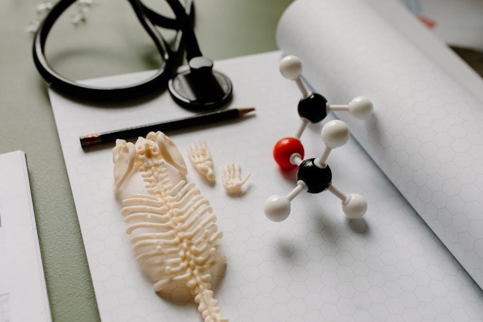
The anatomy and physiology laboratory requires specific tools to facilitate effective learning and experimentation. Essential equipment includes compound microscopes for cellular and tissue analysis, dissection tools like scalpels, forceps, and probes for handling specimens, and measuring instruments such as calipers and rulers for precise measurements. Anatomical models, both physical and digital, are used to visualize complex structures. Storage solutions like specimen containers and slide trays ensure organization and safety. Personal protective equipment (PPE), such as gloves and lab coats, is mandatory for protection during lab activities. Additionally, histology slides, microtome machines, and staining kits are crucial for preparing and examining tissue samples. Modern labs also incorporate digital tools, such as virtual dissection software and 3D anatomy platforms, to enhance understanding. Optional tools like cameras or document cameras can aid in recording observations for further study. These resources collectively create a comprehensive learning environment tailored to anatomy and physiology education.
4.2 Digital Resources and Simulation Software
Digital resources and simulation software are invaluable tools in modern anatomy and physiology education, offering interactive and immersive learning experiences. Platforms like Zygote Body and Visible Body provide detailed 3D models of the human body, enabling students to explore complex structures virtually. Simulation software, such as PhET Interactive Simulations, allows learners to interact with physiological processes like muscle contraction and blood circulation in real-time. Virtual dissection tools, such as Anatomage, replicate the experience of working with cadavers, promoting engagement and understanding. Additionally, online learning platforms integrate quizzes, videos, and interactive modules to reinforce concepts. These resources complement traditional textbooks and lectures, catering to diverse learning styles. Simulation software also supports self-directed learning, enabling students to revisit and review material at their own pace. By leveraging these digital tools, students can deepen their comprehension of anatomy and physiology in a dynamic and accessible manner.
4.3 Effective Use of Microscopy in Anatomy
Microscopy is a cornerstone of anatomy studies, enabling detailed examination of cellular structures that underpin tissue and organ functions. In the anatomy lab, light microscopes are primarily used to study histological slides, while electron microscopes provide higher resolution for intricate cellular details. To use microscopes effectively, students should first prepare slides properly, ensuring accurate staining and mounting. Focusing the lens systematically, starting with low magnification to locate structures before switching to high magnification, is crucial. Proper illumination and use of condensers enhance image clarity. Observing and recording findings methodically helps in correlating microscopic views with anatomical concepts. Regular maintenance of microscopes, including cleaning lenses and adjusting focus, ensures optimal performance. By mastering microscopy techniques, students gain a deeper understanding of cellular anatomy, which is essential for advanced studies in human anatomy and physiology.